
Page 917 - Atlas of Histology with Functional Correlations
P. 917
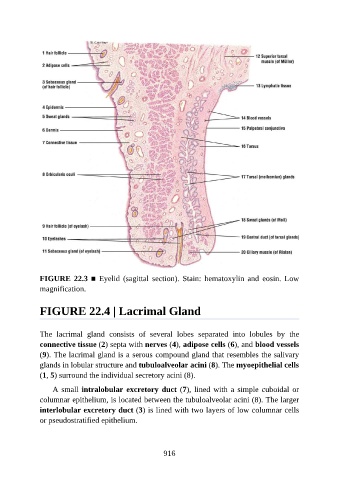
FIGURE 22.3 ■  Eyelid  (sagittal  section).  Stain:  hematoxylin  and  eosin.  Low

               magnification.


               FIGURE 22.4 | Lacrimal Gland


               The  lacrimal  gland  consists  of  several  lobes  separated  into  lobules  by  the
               connective tissue (2) septa with nerves (4), adipose cells (6), and blood vessels
               (9). The lacrimal gland is a serous compound gland that resembles the salivary
               glands in lobular structure and tubuloalveolar acini (8). The myoepithelial cells

               (1, 5) surround the individual secretory acini (8).

                   A small intralobular excretory duct (7), lined with a simple cuboidal or
               columnar epithelium, is located between the tubuloalveolar acini (8). The larger
               interlobular excretory duct (3) is lined with two layers of low columnar cells

               or pseudostratified epithelium.


                                                          916