
Page 922 - Atlas of Histology with Functional Correlations
P. 922
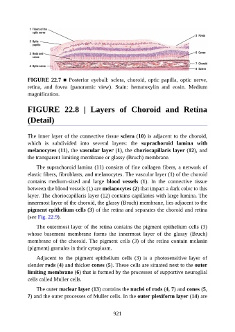
FIGURE  22.7  ■  Posterior  eyeball:  sclera,  choroid,  optic  papilla,  optic  nerve,
               retina,  and  fovea  (panoramic  view).  Stain:  hematoxylin  and  eosin.  Medium
               magnification.


               FIGURE  22.8  |  Layers  of  Choroid  and  Retina

               (Detail)


               The inner layer of the connective tissue sclera (10) is adjacent to the choroid,

               which  is  subdivided  into  several  layers:  the  suprachoroid  lamina  with
               melanocytes (11), the vascular layer (1), the choriocapillaris layer (12), and
               the transparent limiting membrane or glassy (Bruch) membrane.

                   The suprachoroid lamina (11) consists of fine collagen fibers, a network of

               elastic fibers, fibroblasts, and melanocytes. The vascular layer (1) of the choroid
               contains  medium-sized  and  large  blood  vessels  (1).  In  the  connective  tissue
               between the blood vessels (1) are melanocytes (2) that impart a dark color to this
               layer. The choriocapillaris layer (12) contains capillaries with large lumina. The

               innermost layer of the choroid, the glassy (Bruch) membrane, lies adjacent to the
               pigment epithelium cells (3) of the retina and separates the choroid and retina
               (see Fig. 22.9).

                   The outermost layer of the retina contains the pigment epithelium cells (3)

               whose  basement  membrane  forms  the  innermost  layer  of  the  glassy  (Bruch)
               membrane of the choroid. The pigment cells (3) of the retina contain melanin
               (pigment) granules in their cytoplasm.

                   Adjacent  to  the  pigment  epithelium  cells  (3)  is  a  photosensitive  layer  of
               slender rods (4) and thicker cones (5). These cells are situated next to the outer

               limiting membrane (6) that is formed by the processes of supportive neuroglial
               cells called Muller cells.

                   The outer nuclear layer (13) contains the nuclei of rods (4, 7) and cones (5,
               7) and the outer processes of Muller cells. In the outer plexiform layer (14) are


                                                          921