
Page 924 - Atlas of Histology with Functional Correlations
P. 924
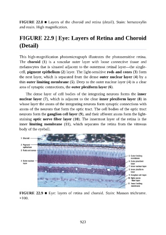
FIGURE 22.8 ■ Layers of the choroid and retina (detail). Stain: hematoxylin

               and eosin. High magnification.


               FIGURE 22.9 | Eye: Layers of Retina and Choroid


               (Detail)


               This  high-magnification  photomicrograph  illustrates  the  photosensitive  retina.
               The  choroid  (1)  is  a  vascular  outer  layer  with  loose  connective  tissue  and
               melanocytes that is situated adjacent to the outermost retinal layer—the single-

               cell, pigment epithelium (2) layer. The light-sensitive rods and cones (3) form
               the next layer, which is separated from the dense outer nuclear layer (4) by a
               thin outer limiting membrane (5). Deep to the outer nuclear layer (4) is a clear
               area of synaptic connections, the outer plexiform layer (6).

                   The  dense  layer  of  cell  bodies  of  the  integrating  neurons  forms  the  inner

               nuclear layer (7), which is adjacent to the clear inner plexiform layer (8) in
               whose layer the axons of the integrating neurons form synaptic connections with
               axons of the neurons that form the optic tract. The cell bodies of the optic tract
               neurons form the ganglion cell layer (9), and their afferent axons form the light-

               staining optic nerve fiber layer (10). The innermost layer of the retina is the
               inner  limiting  membrane  (11),  which  separates  the  retina  from  the  vitreous
               body of the eyeball.


               FIGURE  22.9  ■  Eye:  layers  of  retina  and  choroid.  Stain:  Masson  trichrome.
               ×100.


                                                          923