
Page 928 - Atlas of Histology with Functional Correlations
P. 928
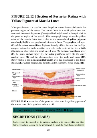
FIGURE 22.12 | Section of Posterior Retina with

               Yellow Pigment of Macula Lutea


               With special stains, it is possible to see the yellow area of the macula lutea in the
               posterior  region  of  the  retina.  The  macula  lutea  is  a  small  yellow  area  that

               surrounds the retinal depression (fovea) and is closely located to the optic disk in
               the  posterior  region  of  the  eyeball.  This  micrograph  image  shows  the  yellow
               color  of  the  macula  lutea  that  is  due  to  the  accumulated  yellow  pigment

               (xanthophyll) (7) in the ganglion cells from the fovea. The ganglion cell layer
               (2) and the retinal axons (1) are displaced laterally off the fovea so that the light
               can pass unimpeded to the sensitive cone cells in the center of the fovea. With
               this stain are also visible the ganglion cell layer (2), the inner plexiform layer
               (8),  the  inner  nuclear  layer  (3),  the  outer  plexiform  layer  (9),  the  outer

               nuclear  layer  (4),  and  the  photoreceptors  cells—the  rods  and  cones  (10).
               Barely visible is the pigment epithelium (5) layer that is adjacent to the dense-
               staining choroid (6). Surrounding the retina is the connective tissue sclera (11).


               FIGURE 22.12 ■ A section of the posterior retina with the yellow pigment of
               the macula lutea. Stain: gold and yellow. ×100.


                  FUNCTIONAL CORRELATIONS 22.1 ■ Eye


                 SECRETIONS (TEARS)


                 Each  eyeball  is  covered  on  its  anterior  surface  with  thin  eyelids  and  fine

                 hairs, eyelashes, located on the margins of the eyelids. Eyelids and eyelashes


                                                          927