
Page 926 - Atlas of Histology with Functional Correlations
P. 926
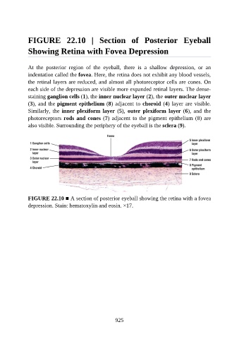
FIGURE  22.10  |  Section  of  Posterior  Eyeball

               Showing Retina with Fovea Depression


               At  the  posterior  region  of  the  eyeball,  there  is  a  shallow  depression,  or  an
               indentation called the fovea. Here, the retina does not exhibit any blood vessels,

               the retinal layers are reduced, and almost all photoreceptor cells are cones. On
               each side of the depression are visible more expanded retinal layers. The dense-
               staining ganglion cells (1), the inner nuclear layer (2), the outer nuclear layer

               (3), and the pigment epithelium (8) adjacent to choroid (4) layer are visible.
               Similarly,  the  inner  plexiform  layer  (5),  outer  plexiform  layer  (6),  and  the
               photoreceptors rods and cones (7) adjacent to the pigment epithelium (8) are
               also visible. Surrounding the periphery of the eyeball is the sclera (9).


               FIGURE 22.10 ■ A section of posterior eyeball showing the retina with a fovea
               depression. Stain: hematoxylin and eosin. ×17.


                                                          925