
Page 919 - Atlas of Histology with Functional Correlations
P. 919
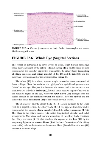
FIGURE  22.5  ■  Cornea  (transverse  section).  Stain:  hematoxylin  and  eosin.
               Medium magnification.


               FIGURE 22.6 | Whole Eye (Sagittal Section)


               The  eyeball  is  surrounded  by  three  layers:  an  outer,  tough  fibrous  connective
               tissue layer composed of the sclera (18) and cornea (1); a middle layer or uvea

               composed of the vascular, pigmented choroid (7), the ciliary body (consisting
               of ciliary processes and ciliary muscle) (4, 14, 15), and the iris (13); and the
               innermost layer composed of the photosensitive retina (8).

                   The  sclera  (18)  is  a  white,  opaque,  tough  connective  tissue  composed  of
               dense collagen fibers that maintain the rigidity of the eyeball and appears as the

               “white”  of  the  eye.  The  junction  between  the  cornea  and  sclera  occurs  at  the
               transition area called the limbus (12), located in the anterior region of the eye. In
               the posterior region of the eye, where the optic nerve (10)  emerges  from  the
               ocular capsule, is the transition between the sclera (18) of the eyeball and the

               connective tissue dura mater (23) of the central nervous system.

                   The choroid (7) and the ciliary body (4, 14, 15) are adjacent to the sclera
               (18). In a sagittal section, the ciliary body (4, 14, 15) appears triangular and is
               composed of the smooth ciliary muscle (14) and the ciliary processes (4, 15).
               The  fibers  in  the  ciliary  muscle  (14)  exhibit  longitudinal,  circular,  and  radial

               arrangements. The folded and vascular extensions of the ciliary body constitute
               the  ciliary  processes  (4,  15)  that  attach  to  the  equator  of  the  lens (16) by the
               suspensory ligament or zonular fibers (5) of the lens. Contraction of the ciliary

               muscle (14) reduces the tension on the zonular fibers (5) and allows the lens (16)
               to assume a convex shape.


                                                          918