
Page 18 - GP Spring 2018
P. 18
A Non-Healing Denture Sore or Something More Sinister?
A Case Report of Oral Histoplasmosis
By David Levenson, DDS, MS and Sonal S. Shah, DDS
Abstract term smokers or those with heavy alcohol Case Report
It is very common for denture patients to consumption. Deep fungal infections, such A 72-year-old male presented to the NYU
experience traumatic sores or ulcers from as histoplasmosis, are typically associated Dental School Urgent Care clinic with a
their dentures, especially with new den- with immunocompromised individuals. chief complaint of a painful denture sore
tures. Such ulcers will usually heal quickly This can be from drugs (immunosuppres- that failed to heal. He said that his new
after adjustment of the denture. If the le- sants, steroids), systemic disease (HIV, lower denture had been adjusted many
sion persists, however, further evaluation leukemia, poorly controlled diabetes) or times over the past six months. The pa-
and biopsy are strongly recommended. An even infancy or old age. Ulcers associated tient’s medical history was noncontributory
1
oral ulcer that did not heal after repeated from denture use are of course associated and his vital signs were normal. He report-
denture adjustments, as in this case report, with the denture. The danger in making ed smoking 5 cigarettes per day for many
became a cause for concern and was even- this last assumption is that valuable time is years.
tually determined to be a Histoplasmosis lost when the diagnosis is wrong; a cancer
deep fungal infection. can metastasize to lymph nodes and a deep Extraoral examination revealed palpable
fungal infection can destroy tissue and dis- and tender right submandibular lymph
Introduction seminate. 2 nodes. Intra-oral examination of his oral
Every dentist cavity revealed an
is familiar with ulcer with an indu-
the classic den- rated border on his
ture sore, an ul- lower right alveo-
cer with a raised lar ridge. It extend-
or hyperplastic ed on the buccal as-
border. They are pect into the right
also familiar with mucobuccal fold
how quickly these area and lingually
sores heal when onto the floor of the
the denture is ad- mouth. The lesion
justed or left out had areas of leuko-
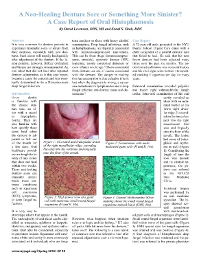
of the mouth for Figure 1: Ulcerated and leukoplakic lesion Figure 2: Granulomas with multi- plakia and erythe-
a few days. Oral of the right mandibular ridge, extending nucleated giant cells (H and E, 10x). ma as well (Figure
healthcare provid- buccally into the vestibule and lingually 1). Considering the
ers are taught to be onto the floor of the mouth. length of time the
wary of any lesion sore was present
that does not heal and its clinical ap-
within two weeks. pearance, the pa-
These non-healing tient was referred
denture sores can to the NYUCD
clinically mimic Oral Medicine
much more om- Clinic.
inous conditions
such as squamous Incisional biopsy
cell carcinoma was performed by
(SCC), lymphoma, Figure 3: High power view of a giant Figure 4: Gomori Methenamine Silver an oral medicine
or deep fungal in- cell with numerous small round fungal staining shows the small round fungal specialist. The bi-
fections. organisms (H and E, 40x). organisms stained black (GMS, 40x). opsy showed sev-
eral granulomas
It is very easy to with multinucleat-
stereotype ulcers that appear in the mouth. ed giant cells and macrophages (Figure 2).
The vast majority of oral ulcers can be clas- However, what happens when intuition Small round fungal organisms were identi-
sified as traumatic, aphthous or herpetic. rules over logic and the feeling, “ If I take fied within some of the giant cells (Figure
However, malignancy and systemic infec- off just a little bit more from the denture,” 3). GMS special stain for fungal organisms
tions must also be considered, especially takes over? The following is a case report was ordered and was positive (Figure 4).
in persistent lesions. Squamous cell carci- of a denture sore that refused to heal after A final diagnosis of histoplasmosis deep
noma of the oral cavity is more commonly repeated adjustments over a six-month pe- fungal infection was rendered and the pa-
associated with individuals who are long- riod. tient was referred to his private physician
www.nysagd.org l Spring 2018 l GP 18