Page 4 - Prueba
P. 4

2        Eyes and vision


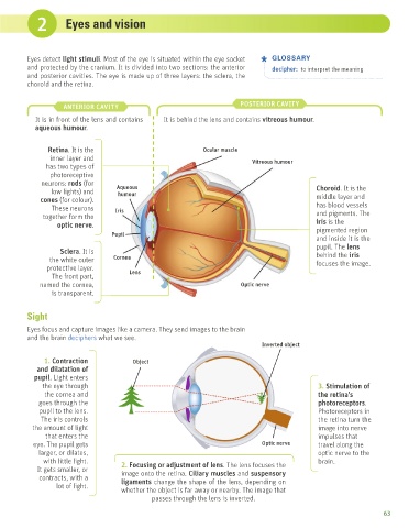
               Eyes detect light stimuli. Most of the eye is situated within the eye socket   GLOSSARY
               and protected by the cranium. It is divided into two sections: the anterior   decipher:  to interpret the meaning
               and posterior cavities. The eye is made up of three layers: the sclera, the
               choroid and the retina.


                          ANTERIOR CAVITY                                        POSTERIOR CAVITY
                 It is in front of the lens and contains   It is behind the lens and contains vitreous humour.
                 aqueous humour.


                     Retina. It is the                               Ocular muscle
                      inner layer and                                                Vitreous humour
                    has two types of
                      photoreceptive
                   neurons: rods (for     Aqueous
                      low lights) and      humour                                                        Choroid. It is the
                   cones (for colour).                                                                   middle layer and
                      These neurons       Iris                                                           has blood vessels
                   together form the                                                                     and pigments. The
                        optic nerve.                                                                     iris is the
                                         Pupil                                                           pigmented region
                                                                                                         and inside it is the
                                                                                                         pupil. The lens
                         Sclera. It is                                                                   behind the iris
                     the white outer     Cornea                                                          focuses the image.
                     protective layer.        Lens
                      The front part,
                  named the cornea,                                              Optic nerve
                      is transparent.


               Sight

               Eyes focus and capture images like a camera. They send images to the brain
               and the brain deciphers what we see.
                                                                                        Inverted object

                    1. Contraction             Object
                  and dilatation of
                 pupil. Light enters
                   the eye through                                                                       3. Stimulation of
                    the cornea and                                                                       the retina’s
                  goes through the                                                                       photoreceptors.
                  pupil to the lens.                                                                     Photoreceptors in
                   The iris controls                                                                     the retina turn the
                the amount of light                                                                      image into nerve
                    that enters the                                                                      impulses that
                eye. The pupil gets                                                     Optic nerve      travel along the
                  larger, or dilates,                                                                    optic nerve to the
                    with little light.      2. Focusing or adjustment of lens. The lens focuses the      brain.
                  It gets smaller, or       image onto the retina. Ciliary muscles and suspensory
                  contracts, with a         ligaments change the shape of the lens, depending on
                        lot of light.
                                            whether the object is far away or nearby. The image that
                                                     passes through the lens is inverted.

                                                                                                                              63
   1   2   3   4   5   6   7   8   9