
Page 12 - DP Vol 20 No 5_Neat
P. 12
COSMETIC SECTION
STRATEGIC APPROACHES TO ADDRESSING
ADJACENT CLASS 2 DEFECTS IN
QUADRANT DENTISTRY
Anand Narvekar
INTRODUCTION
Losing any of the tooth contacts disrupts the continuity of the enamel
layer, significantly reducing tooth rigidity. The strength of a tooth
decreases proportionally to the amount of tooth tissue removed.
Specifically, the loss of one contact can reduce tooth strength by over
35%, while the loss of both contacts can reduce strength by more than
65%. This reduction is attributed to the high volume of enamel in the
proximal areas. For clinicians, success is achieved by ensuring well-
sealed restorations with proper contacts and contours. This article
offers a practical and reliable approach to managing adjacent multiple
Class II direct resin restorations in routine clinical practice.
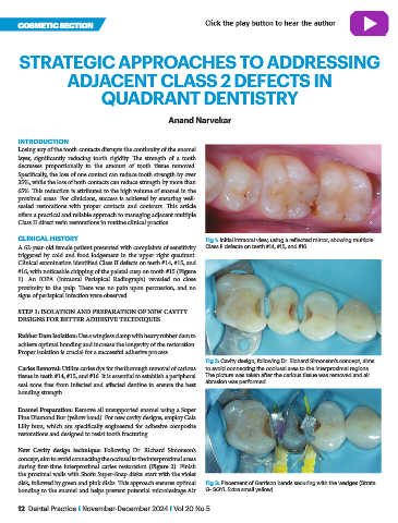
CLINICAL HISTORY Fig 1: Initial intraoral view, using a reflected mirror, showing multiple
A 62-year-old female patient presented with complaints of sensitivity Class II defects on teeth #14, #15, and #16.
triggered by cold and food lodgement in the upper right quadrant.
Clinical examination identified Class II defects on teeth #14, #15, and
#16, with noticeable chipping of the palatal cusp on tooth #15 (Figure
1). An IOPA (Intraoral Periapical Radiograph) revealed no close
proximity to the pulp. There was no pain upon percussion, and no
signs of periapical infection were observed.
STEP 1: ISOLATION AND PREPARATION OF NEW CAVITY
DESIGNS FOR BETTER ADHESIVE TECHNIQUES
Rubber Dam Isolation: Use a wingless clamp with heavy rubber dam to
achieve optimal bonding and increase the longevity of the restoration.
Proper isolation is crucial for a successful adhesive process.
Fig 2: Cavity design, following Dr. Richard Simonson’s concept, aims
Caries Removal: Utilize caries dye for the thorough removal of carious to avoid connecting the occlusal area to the interproximal regions.
tissue in teeth #14, #15, and #16. It is essential to establish a peripheral The picture was taken after the carious tissue was removed and air
seal zone free from infected and affected dentine to ensure the best abrasion was performed.
bonding strength.
Enamel Preparation: Remove all unsupported enamel using a Super
Fine Diamond Bur (yellow band). For new cavity designs, employ Cala
Lilly burs, which are specifically engineered for adhesive composite
restorations and designed to resist tooth fracturing.
New Cavity design technique: Following Dr. Richard Simonson’s
concept, aim to avoid connecting the occlusal to the interproximal areas
during first-time interproximal caries restoration (Figure 2). Finish
the proximal walls with Shofu Super-Snap disks: start with the violet
disk, followed by green and pink disks. This approach ensures optimal Fig 3: Placement of Garrison bands securing with the wedges (Strata
bonding to the enamel and helps prevent potential microleakage.Air G- SGYL Extra small yellow)
12 Dental Practice I November-December 2024 I Vol 20 No 5