
Page 16 - DP Vol 20 No 5_Neat
P. 16
PERIODONTIC SECTION
AN IMPROVED PERI-IMPLANTITIS OUTLOOK
Senthil Thiagarajan describes decontamination of the implant surface and surrounding tissue
for a patient suffering from peri-implantitis
Peri-implantitis is a destructive inflammatory process that may be caused by
occlusion, loose implant abutment, smoking or inadequate oral hygiene resulting
in plaque.
The following case highlights the versatility of erbium YAG laser technology, as
part of an effective treatment protocol for serious dental conditions.
When planning cases, as clinicians, we often turn to tried-and-tested products,
materials and equipment for reliability and predictability, both in the procedure,
as well as in the final outcome.
Within this context, laser technology is becoming increasingly indispensable
for treatment of a wide range of clinical indications. Used appropriately, it can lead
to safer, faster and more predictable outcomes and a more comfortable experience
for the patient.
CASE SUMMARY
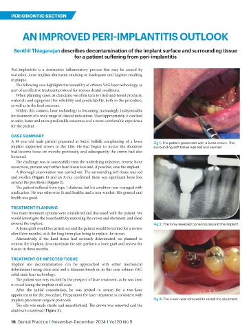
A 60-year-old male patient presented at Smile Suffolk complaining of a loose Fig 1: The patient presented with a loose crown. The
implant supported crown in the LR6. He had begun to notice the abutment surrounding soft tissue was red and swollen
had become loose six months previously, and subsequently the crown had also
loosened.
The challenge was to successfully treat the underlying infection, reverse bone
resorption, prevent any further hard tissue loss and, if possible, save the implant.
A thorough examination was carried out. The surrounding soft tissue was red
and swollen (Figure 1) and an X-ray confirmed there was significant bone loss
around the prosthesis (Figure 2).
The patient suffered from type 1 diabetes, but his condition was managed with
medication. He was otherwise fit and healthy and a non-smoker. His general oral
health was good.
TREATMENT PLANNING
Two main treatment options were considered and discussed with the patient. We
would investigate the bone health by removing the crown and abutment, and clean
around the implant. Fig 2: The X-ray revealed bone loss around the implant
A bone graft would be carried out and the patient would be invited for a review
after three months, with the long-term plan being to replace the crown.
Alternatively, if the hard tissue had seriously deteriorated, we planned to
remove the implant, decontaminate the site, perform a bone graft and review the
tissues in three months.
TREATMENT OF INFECTED TISSUE
Implant site decontamination can be approached with either mechanical
debridement using citric acid and a titanium brush or, in this case, erbium YAG
solid-state laser technology.
The patient was very excited by the prospect of laser treatment, as he was keen
to avoid losing the implant at all costs.
After the initial consultation, he was invited to return for a two-hour
appointment for the procedure. Preparation for laser treatment is consistent with
implant placement surgical protocols. Fig 3: The crown was removed to reveal the abutment
The site was made sterile and anaesthetised. The crown was removed and the
abutment examined (Figure 3).
16 Dental Practice I November-December 2024 I Vol 20 No 5