
Page 60 - Power of Stem Cells- arthritis and regeneration
P. 60
Theranostics 2018, Vol. 8, Issue 4 914
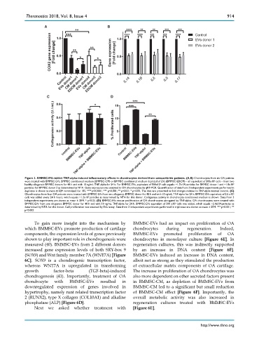
Figure 3. BMMSC-EVs inhibit TNF-alpha-induced inflammatory effects in chondrocytes derived from osteoarthritic patients. (A,B) Chondrocytes from six OA patients
were treated with BMMSC-EVs, BMMSC conditioned medium (BMMSC-CM) or BMMSC conditioned medium depleted of EVs (BMMSC-EDCM) – all equivalent of 500x10 3 cells – from two
healthy allogeneic BMMSC donors for 48 h and with 10 ng/mL TNF-alpha for 24 h. For BMMSC-EVs, equivalent of 500x10 3 cells equals: ~1.7x10 8 particles for BMMSC donor 1 and ~1.8x10 9
particles for BMMSC donor 2 as determined by NTA. Gene expression was analyzed in OA chondrocytes by qRT-PCR. Quantification of data from 3 independent experiments performed in
duplicate is shown as mean ± SEM normalized for 18S. ****p<0.0001; *** p<0.001; ** p<0.01; * p<0.05. The data are presented as fold changes relative to TNF-alpha treated control. (C)
Chondrocytes from four OA patients were treated with BMMSC-EVs from one allogeneic BMMSC donor for 48 h and with 10 ng/mL TNF-alpha for 24 h. BMMSC-EVs equivalent of 2.5 x10 6
cells was added every 24 h hours, which equals ~1.2x10 9 particles as determined by NTA for this donor. Collagenase activity in chondrocyte conditioned medium is shown. Data from 3
independent experiments are shown as mean ± SEM. * p<0.05. (D) BMMSC-EVs rescue proliferation of OA chondrocytes abrogated by TNF-alpha. OA chondrocytes were treated with
BMMSC-EVs from one allogeneic BMMSC donor for 48 h and with 10 ng/mL TNF-alpha for 24 h. BMMSC-EVs equivalent of 500 x10 3 cells was added, which equals ~2.4x10 8 particles as
determined by NTA for this donor. Cell proliferation was assessed by EdU assay. Data from 2 independent experiments performed in triplicates are shown as mean ± SEM. *** p<0.001; **
p<0.002
To gain more insight into the mechanism by BMMSC-EVs had an impact on proliferation of OA
which BMMSC-EVs promote production of cartilage chondrocytes during regeneration. Indeed,
components, the expression levels of genes previously BMMSC-EVs promoted proliferation of OA
shown to play important role in chondrogenesis were chondrocytes in monolayer culture [Figure 6E]. In
measured (40). BMMSC-EVs from 2 different donors regeneration cultures, this was indirectly supported
increased gene expression levels of both SRY-box 9 by an increase in DNA content [Figure 6F].
(SOX9) and Wnt family member 7A (WNT7A) [Figure BMMSC-EVs induced an increase in DNA content,
6C]. SOX9 is a chondrogenic transcription factor, albeit not as strong as they stimulated the production
whereas WNT7A is upregulated in transforming of extracellular matrix components of OA cartilage.
growth factor-beta (TGF-beta)-induced The increase in proliferation of OA chondrocytes was
chondrogenesis (41). Importantly, treatment of OA also more dependent on other secreted factors present
chondrocyte with BMMSC-EVs resulted in in BMMSC-CM, as depletion of BMMSC-EVs from
downregulated expression of genes involved in BMMSC-CM led to a significant but small reduction
hypertrophy, namely runt related transcription factor of BMMSC-CM effect [Figure 6F]. Importantly, the
2 (RUNX2), type X collagen (COL10A1) and alkaline overall metabolic activity was also increased in
phosphatase (ALP) [Figure 6D]. regeneration cultures treated with BMMSC-EVs
Next we asked whether treatment with [Figure 6E].
http://www.thno.org