
Page 57 - Power of Stem Cells- arthritis and regeneration
P. 57
Theranostics 2018, Vol. 8, Issue 4 911
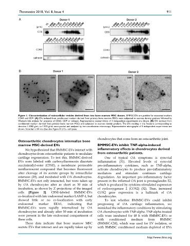
Figure 1. Characterization of extracellular vesicles derived from two bone marrow MSC donors. BMMSC-EVs are positive for exosomal markers
CD63 and CD9. (A) EVs isolated from conditioned medium derived from primary bone marrow MSCs were subjected to sucrose density gradient followed by
Western blot analysis for presence of CD63, CD9 or calnexin. Representative western-blots of 3 independent experiments are shown. (B) EVs isolated from
conditioned medium derived from primary bone marrow MSCs and subjected to sucrose density gradient. The EVs residing in the fractions corresponding to
densities 1.1082 g/mL to 1.972 g/mL were pooled and analyzed by immuno-electron microscopy. Representative micrographs of 3 independent experiments are
shown. Scale bar is 50 nm. (See also Figure S1.) CL- cell lysate.
chondrocytes that come from an osteoarthritic joint.
Osteoarthritic chondrocytes internalize bone
marrow MSC-derived EVs BMMSC-EVs inhibit TNF-alpha-induced
We hypothesized that BMMSC-EVs interact with inflammatory effects in chondrocytes derived
chondrocytes from osteoarthritic patients to modulate from osteoarthritic patients.
cartilage regeneration. To test this, BMMSC-derived One of typical OA symptoms is synovial
EVs were labeled with carboxyfluorescein diacetate inflammation (31). Elevated levels of synovial
succinimidyl-ester (CFSE), a membrane permeable pro-inflammatory cytokines, such as TNF-alpha,
nonfluorescent compound that becomes fluorescent activate chondrocytes to produce pro-inflammatory
after cleavage of its acetate groups by intracellular mediators and stimulate continues cartilage
esterases (29), and incubated with OA chondrocytes. degradation. An important pro-inflammatory factor
BMMSC-EVs not only interacted, but were taken up present in the inflamed OA joint is prostaglandin E2,
by OA chondrocytes after as short as 30 min of which is produced by cytokine-stimulated expression
incubation, as shown by Z projections of the imaged of cyclooxygenase 2 (COX2) (32). Thus, increased
cells [Figure 2]. CFSE-labeled BMMSC-EVs COX2 gene expression is a hallmark of OA
co-localized with late endosomal marker LAMP-1, but chondrocytes.
showed little or no co-localization with early To test whether BMMSC-EVs could inhibit
endosomal marker EEA1, indicating that progressing of OA cartilage inflammation, we
BMMSC-EVs were rapidly internalized by OA mimicked the inflammatory conditions by stimulating
chondrocytes and already after 30 min of incubation OA chondrocytes with TNF-alpha. TNF-alpha treated
were present in the late endosomal compartment of cells were incubated for 48 h with BMMSC-EVs or
these cells. with conditioned medium from BMMSC
These data indicate that bone marrow MSC (BMMSC-CM), which was used for EV isolation, or
secrete EVs that interact and are rapidly taken up by with BMMSC conditioned medium depleted of EVs
http://www.thno.org