
Page 61 - Power of Stem Cells- arthritis and regeneration
P. 61
Theranostics 2018, Vol. 8, Issue 4 915
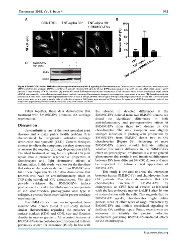
Figure 4. BMMSC-EVs inhibit TNF-alpha-induced pro-inflammatory NFκB signaling in OA chondrocytes. Chondrocytes from two OA patients were treated with
BMMSC-EVs from one allogeneic BMMSC donor for 24 h and with 10 ng/mL TNF-alpha for 30 min. BMMSC-EVs equivalent of 2.5 x10 6 cells was added, which equals ~1.2x10 9
particles as determined by NTA for this donor. (A) BMMSC-EVs inhibit TNF-alpha-induced nuclear translocation of p65 subunit of NFκB. Nuclear translocation of p65 subunit
of NFκB was assessed by immunofluorescence analysis and confocal microscopy. Representative images of two independent experiments are shown. (B) Quantification of data
presented in A. Data from 2 independent experiments are shown as mean ± SEM. (C) BMMSC-EVs abrogate TNF-alpha-induced phosphorylation of IκBα. The OA chondrocytes
were treated as in (A) and lysed in sample buffer directly after treatment. Cell lysates were analyzed by Western-blot for presence of pIκBα. Representative results of two
independent experiments performed with chondrocytes of four OA donors are shown.
Taken together, these data demonstrate that the absence of detected differences in the
treatment with BMMSC-EVs promotes OA cartilage BMMSC-EVs derived from two BMMSC donors, we
regeneration. found no significant differences in both
anti-inflammatory and pro-regenerative effects of
Discussion BMMSC-EVs from these two donors on OA
Osteoarthritis is one of the most prevalent joint chondrocytes. The only exception was slightly
diseases and a major public health problem. It is stronger induction of proteoglycan production by
characterized by progressive articular cartilage BMMSC-EVs from BMMSC donor two in OA
destruction and synovitis (42,43). Current therapies chondrocytes [Figure 5B]. Screening of more
attempt to relieve the symptoms, but they cannot stop BMMSC-EVs donors should facilitate defining
or reverse the ongoing cartilage degeneration (4,44). whether this minor difference in the BMMSC-EVs
The ideal treatment aiming for an optimal OA joint effect on proteoglycan production is a more general
repair should promote regenerative properties of phenomenon that results in real functional differences
chondrocytes and fight destructive effects of between EVs from different BMMSC donors and may
inflammation. In this study we show for the first time be important for future clinical application of
that extracellular vesicles derived from BMMSC may BMMSC-EVs.
fulfil these requirements. Our data demonstrate that This study is the first to show the interaction
BMMSC-EVs have an anti-inflammatory effect on between human BMMSC-EVs and chondrocytes from
TNF-alpha-stimulated OA chondrocytes. We also OA patients. Our data demonstrate that OA
provide evidence that BMMSC-EVs induce chondrocytes internalize BMMSC-EVs by
production of crucial extracellular matrix components endocytosis, as CFSE labeled vesicles co-localized
of OA chondrocytes, proteoglycans and type II with the late endocytic marker LAMP-1 after 30 min
collagen, a process that is essential for proper cartilage of co-incubation with the cells. This suggests that by
regeneration. BMMSC-EV uptake, chondrocytes might utilize
The BMMSC-EVs from two independent bone protein, RNA or other types of cargo transferred by
marrow MSC donors tested in our study showed BMMSC-EVs and initiate specialized signaling to
similar characteristics regarding the presence of facilitate OA cartilage repair. Future experiments are
surface markers (CD63 and CD9), size and flotation necessary to identify the precise molecular
density in sucrose gradient. All reported features of mechanism governing BMMSC-EV-mediated effects
BMMSC-EVs from both donors corresponded to those on OA chondrocytes.
previously shown for exosomes (45–47). In line with
http://www.thno.org