
Page 82 - Human Umbilical Cord Mesenchymal Stem Cells
P. 82
1482 R. Friedman et al.
A 60
38.4%
50 p = 0.001
% human CD45+ cells 30 p = 0.02
40
20
10
0 1.9% 6.7% 5.9%
MNC MNC + UC-MSC MNC MNC + UC-MSC
Marrow Peripheral Blood
B 20
18 p = 0.03
16
% human CD45+ cells 12 8 6 4.5% 10.1% p = 0.57
14
10
2 4 2.7% 2.1%
0
CD 34+ cells CD 34+ cells + CD 34+ cells CD 34+ cells +
UC-MSC UC-MSC
Marrow Peripheral Blood
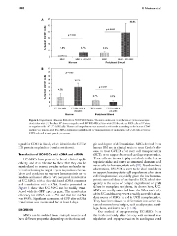
Figure 2. Engraftment of human BM cells in NOD/SCID mice. The mice underwent transplantation (intravenous injec-
6
4
6
tion) either with UCB cells at 10 alone or together with 10 UC-MSCs (A) or with CD34-enriched UCB cells at 10 alone
6
or together with 10 UC-MSCs (B). Human cell engraftment was assessed at 6-8 weeks according to the human CD45
marker. Co-transplanted UC-MSCs augmented engraftment for transplantation of unfractionated UCB cells as well as
CD34 selected hematopoietic precursors.
signal for CD41 in blood, which identifies the GPIIa/ gin and degree of differentiation. MSCs derived from
IIIb protein on platelets (results not shown). human BM are in clinical trials to treat Crohn’s dis-
ease, to treat GVHD after stem cell transplantation
Transfection of UC-MSCs with cDNA and mRNA (SCT), or to support bone and cartilage regeneration.
UC-MSCs have potentially broad clinical appli- These cells are known to play a vital role in the hema-
cability, and it is relevant to show that they can be topoietic niche and serve as structural elements and
manipulated to express certain surface molecules in- nurse cells for hematopoietic cells [26]. Based on these
volved in homing to target organs to produce chemo- observations, BM-MSCs seem to be ideal candidates
kines and cytokines to support hematopoiesis or to to support hematopoietic cell engraftment after stem
mediate antitumor effects. We compared transfection cell transplantation, especially given the low hemato-
of UC-MSCs with a plasmid-based cDNA construct poietic stem cell dose often found in UCB, which fre-
and transfection with mRNA. Results presented in quently is the cause of delayed engraftment or graft
Figure 3 show that UC-MSC can be readily trans- failure in transplant recipients. As shown here, UC-
fected with the GFP reporter gene. The transfection MSCs are readily extracted from the Wharton’s jelly
efficiency for cDNA was 30.9% and that for mRNA of the UC and thus represent a readily accessible abun-
was 89.8%. Significant expression of GFP after mRNA dant source of MSCs to aid in UCB transplantation.
They have been shown to differentiate into other tis-
transfection was maintained for at least 4 days.
sues of mesenchymal origin, such as adipocytes, carti-
lage, bone, and nerve cells [14-16].
DISCUSSION
Our method of cryopreserving UC-MSCs from
MSCs can be isolated from multiple sources and the fresh cord early after delivery with minimal ma-
have different properties depending on the tissue ori- nipulation and cryopreservation in autologous cord