
Page 95 - Human Umbilical Cord Mesenchymal Stem Cells
P. 95
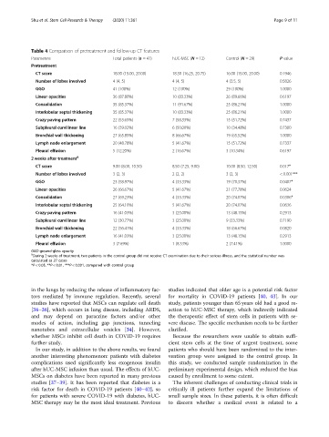
Shu et al. Stem Cell Research & Therapy          (2020) 11:361                          Page 9 of 11


            Table 4 Comparison of pretreatment and follow-up CT features
            Parameters                    Total patients (n = 41)  hUC-MSC (N = 12)  Control (N = 29)  P value
            Pretreatment
              CT score                    18.00 (15.00, 20.00)  18.50 (16.25, 20.75)  16.00 (15.00, 20.00)  0.1946
              Number of lobes involved    4 (4, 5)             4 (4, 5)            4 (3.5, 5)         0.5826
              GGO                         41 (100%)            12 (100%)           29 (100%)          1.0000
              Linear opacities            36 (87.80%)          10 (83.33%)         26 (89.66%)        0.6197
              Consolidation               35 (85.37%)          11 (91.67%)         25 (86.21%)        1.0000
              Interlobular septal thickening  35 (85.37%)      10 (83.33%)         25 (86.21%)        1.0000
              Crazy-paving pattern        22 (53.65%)          7 (58.33%)          15 (51.72%)        0.7437
              Subpleural curvilinear line  16 (39.02%)         6 (50.00%)          10 (34.48%)        0.7300
              Bronchial wall thickening   27 (65.85%)          8 (66.67%)          19 (65.52%)        1.0000
              Lymph node enlargement      20 (48.78%)          5 (41.67%)          15 (51.72%)        0.7337
              Pleural effusion            5 (12.20%)           2 (16.67%)          3 (10.34%)         0.6197
            2 weeks after treatment §
              CT score                    9.00 (8.00, 10.50)   8.50 (7.25, 9.00)   10.00 (8.50, 12.50)  0.017*
              Number of lobes involved    3 (2, 3)             2 (2, 2)            3 (2, 3)           < 0.001***
              GGO                         23 (58.97%)          4 (33.33%)          19 (70.37%)        0.0407*
              Linear opacities            26 (66.67%)          5 (41.67%)          21 (77.78%)        0.0624
              Consolidation               27 (69.23%)          4 (33.33%)          20 (74.07%)        0.0306*
              Interlobular septal thickening  25 (64.10%)      5 (41.67%)          20 (74.07%)        0.0636
              Crazy-paving pattern        16 (41.03%)          3 (25.00%)          13 (48.15%)        0.2913
              Subpleural curvilinear line  12 (30.77%)         3 (25.00%)          9 (33.33%)         0.7190
              Bronchial wall thickening   22 (56.41%)          4 (33.33%)          18 (66.67%)        0.0820
              Lymph node enlargement      16 (41.03%)          3 (25.00%)          13 (48.15%)        0.2913
              Pleural effusion            3 (7.69%)            1 (8.33%)           2 (7.41%)          1.0000
            GGO ground-glass opacity
            §
             During 2 weeks of treatment, two patients in the control group did not receive CT examination due to their serious illness, and the statistical number was
            calculated as 27 cases
            *P < 0.05, **P < 0.01, ***P < 0.001, compared with control group


            in the lungs by reducing the release of inflammatory fac-  studies indicated that older age is a potential risk factor
            tors mediated by immune regulation. Recently, several  for mortality in COVID-19 patients [40, 43]. In our
            studies have reported that MSCs can regulate cell death  study, patients younger than 65 years old had a good re-
            [34–36], which occurs in lung disease, including ARDS,  action to hUC-MSC therapy, which indirectly indicated
            and may depend on paracrine factors and/or other  the therapeutic effect of stem cells in patients with se-
            modes of action, including gap junctions, tunneling  vere disease. The specific mechanism needs to be further
            nanotubes and extracellular vesicles [34]. However,  clarified.
            whether MSCs inhibit cell death in COVID-19 requires  Because the researchers were unable to obtain suffi-
            further study.                                    cient stem cells at the time of urgent treatment, some
              In our study, in addition to the above results, we found  patients who should have been randomized to the inter-
            another interesting phenomenon: patients with diabetes  vention group were assigned to the control group. In
            complications used significantly less exogenous insulin  this study, we conducted sample randomization in the
            after hUC-MSC infusion than usual. The effects of hUC-  preliminary experimental design, which reduced the bias
            MSCs on diabetes have been reported in many previous  caused by enrollment to some extent.
            studies [37–39]. It has been reported that diabetes is a  The inherent challenges of conducting clinical trials in
            risk factor for death in COVID-19 patients [40–42], so  critically ill patients further expand the limitations of
            for patients with severe COVID-19 with diabetes, hUC-  small sample sizes. In these patients, it is often difficult
            MSC therapy may be the most ideal treatment. Previous  to discern whether a medical event is related to a