
Page 285 - Atlas of Small Animal CT and MRI
P. 285
Cranial Nerves 275
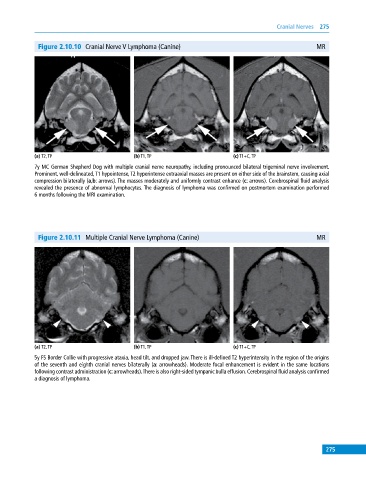
Figure 2.10.10 Cranial Nerve V Lymphoma (Canine) MR
(a) T2, TP (b) T1, TP (c) T1+C, TP
7y MC German Shepherd Dog with multiple cranial nerve neuropathy, including pronounced bilateral trigeminal nerve involvement.
Prominent, well‐delineated, T1 hypointense, T2 hyperintense extraaxial masses are present on either side of the brainstem, causing axial
compression bilaterally (a,b: arrows). The masses moderately and uniformly contrast enhance (c: arrows). Cerebrospinal fluid analysis
revealed the presence of abnormal lymphocytes. The diagnosis of lymphoma was confirmed on postmortem examination performed
6 months following the MRI examination.
Figure 2.10.11 Multiple Cranial Nerve Lymphoma (Canine) MR
(a) T2, TP (b) T1, TP (c) T1+C, TP
5y FS Border Collie with progressive ataxia, head tilt, and dropped jaw. There is ill‐defined T2 hyperintensity in the region of the origins
of the seventh and eighth cranial nerves bilaterally (a: arrowheads). Moderate focal enhancement is evident in the same locations
following contrast administration (c: arrowheads). There is also right‐sided tympanic bulla effusion. Cerebrospinal fluid analysis confirmed
a diagnosis of lymphoma.
275