
Page 282 - Atlas of Small Animal CT and MRI
P. 282
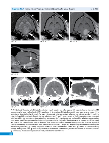
Figure 2.10.7 Cranial Nerve V Benign Peripheral Nerve Sheath Tumor (Canine) CT & MR
(a) T2, TP (b) T1, TP (c) T1+C, TP
(d) T1+C, SP
(e) CT, TP (f) CT+C, TP (g) CT+C, SP
7y MC Shetland Sheepdog with left‐sided masticatory muscle atrophy and other signs of left trigeminal nerve dysfunction. MR
images reveal a large, well‐delineated, T1 hypointense, T2 hyperintense left‐sided extraaxial mass causing compression of the
brainstem and cerebellum (a–d: arrow). The mass intensely and uniformly contrast enhances and extends rostrally through the
trigeminal canal (d: arrowhead). There is also marked atrophy and T1 and T2 hyperintensity of the left masseter muscle, consistent
with fatty infiltration from chronic denervation (a,b: arrowheads). A CT examination was performed for radiation treatment plan
ning. Images e and f are unenhanced and enhanced images acquired at the level of the trigeminal canal. Images h and i (next page)
are more caudal, centered at the level of the mass. There is destruction of the temporal bone pyramid that forms the trigeminal
canal (e: arrowhead). Note that the mass is not seen on the unenhanced CT images (h). The previously identified mass intensely but
nonuniformly enhances (f,g,i: arrow). The globoid part of the mass is located in the caudal fossa (g,i: arrow) but extends rostrally
through the trigeminal canal (g: arrowhead). Postmortem examination confirmed the presence and location of the extraaxial mass
(j: arrowheads). Microscopic diagnosis was left trigeminal nerve neurofibroma.
272