
Page 283 - Atlas of Small Animal CT and MRI
P. 283
Cranial Nerves 273
Figure 2.10.7 (Continued ) CT & MR
(h) CT, TP (i) CT+C, TP (j) GP, VENT
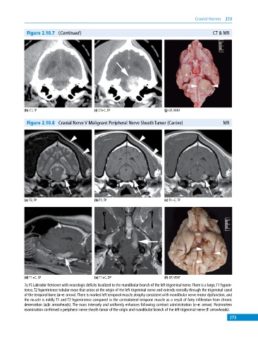
Figure 2.10.8 Cranial Nerve V Malignant Peripheral Nerve Sheath Tumor (Canine) MR
(a) T2, TP (b) T1, TP (c) T1+C, TP
(d) T1+C, SP (e) T1+C, DP (f) GP, VENT
7y FS Labrador Retriever with neurologic deficits localized to the mandibular branch of the left trigeminal nerve. There is a large, T1 hypoin
tense, T2 hyperintense tubular mass that arises at the origin of the left trigeminal nerve and extends rostrally through the trigeminal canal
of the temporal bone (a–e: arrow). There is marked left temporal muscle atrophy consistent with mandibular nerve motor dysfunction, and
the muscle is mildly T1 and T2 hyperintense compared to the contralateral temporal muscle as a result of fatty infiltration from chronic
denervation (a,b: arrowheads). The mass intensely and uniformly enhances following contrast administration (c–e: arrow). Postmortem
examination confirmed a peripheral nerve sheath tumor of the origin and mandibular branch of the left trigeminal nerve (f: arrowheads).
273