
Page 284 - Atlas of Small Animal CT and MRI
P. 284
274 Atlas of Small Animal CT and MRI
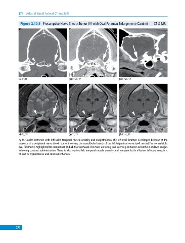
Figure 2.10.9 Presumptive Nerve Sheath Tumor (V) with Oval Foramen Enlargement (Canine) CT & MR
(a) CT, TP (b) CT+C, TP (c) CT+C, SP
(d) T2, TP (e) T1, TP (f) T1+C, TP
7y FS Golden Retriever with left‐sided temporal muscle atrophy and enophthalmos. The left oval foramen is enlarged because of the
presence of a peripheral nerve sheath tumor involving the mandibular branch of the left trigeminal nerve. (a–f: arrow) The normal right
oval foramen is highlighted for comparison (a,b,d–f: arrowhead). The mass uniformly and intensely enhances on both CT and MR images
following contrast administration. There is also marked left temporal muscle atrophy and tympanic bulla effusion. Affected muscle is
T1 and T2 hyperintense and contrast enhances.
274