
Page 279 - Atlas of Small Animal CT and MRI
P. 279
Cranial Nerves 269
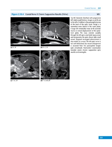
Figure 2.10.4 Cranial Nerve II Chronic Suppurative Neuritis (Feline) MR
12y MC Domestic Shorthair with progressive
left‐sided exophthalmos. Images a and b are
to the left of midline in the parasagittal plane
at the level of the optic canal. Image c is
somewhat more lateral in the same plane.
A large left retrobulbar mass (a–d: asterisk)
results in rostral displacement of the ipsila
teral globe. The mass extends caudally
through the left optic canal (a,b: large arrow)
and incorporates the optic chiasm (a,b: small
arrow). Regional meningeal enhancement is
also evident (c: arrow). The left optic nerve is
not well delineated, but neural enlargement
is assumed from the parasagittal images
(a) T1, SP (b) T1+C, SP (a,b: arrowhead). Postmortem examination
revealed severe chronic suppurative optic
neuritis and meningitis.
(c) T1+C, SP (d) T1+C+FS, DP
269