
Page 294 - Atlas of Small Animal CT and MRI
P. 294
284 Atlas of Small Animal CT and MRI
the internal extent and character of the sinus, with Vascular anomalies
increasing grade reflecting a more invasive lesion.
38
Of the few reported cases, most are at the level of the Although vascular anomalies involving the spinal cord are
cervical or cranial thoracic vertebral column. Dermoid rare in dogs and cats, various disorders have been sporadi
sinuses have been reported in dogs and cats, and cally reported as single case reports or small case studies.
Rhodesian Ridgebacks are predisposed. 39–43 Abnormalities have included arterial malformation,
CT imaging features may be unremarkable other than a arteriovenous malformation, hemangioma, intramedul
possible skin surface defect. Conventional radiographic lary cavernous malformation, and hamartoma. Intrinsic
sinusography has been used to determine the internal vascular anomalies can displace spinal cord parenchyma,
margin of the sinus and assess its association to the under and both intrinsic and extrinsic anomalies can cause cord
46–51
lying vertebral column, meninges, and spinal cord, and compression.
CT would likely be of similar value. Reported MR imag CT and MR imaging features are dependent on the
ing features include superficial mass with mixed T1 inten type and location of the anomaly. Large vessel anomalies
sity, mild T2 hyperintensity, and STIR hyperintensity. In can clearly be seen on contrast‐enhanced imaging
some instances, a sinus tract was not seen, and the depth studies (Figure 3.1.16). Imaging signs of intrinsic spinal
of the lesion was underestimated. In one other patient, a cord disease can be seen with intramedullary lesions,
T2 hyperintense tract was clearly identified coursing from and T2* sequences may be helpful in characterizing
the superficial mass to the dura matter. 2,44,45 thrombotic disease.
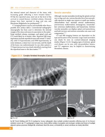
Figure 3.1.1 Complex Vertebral Anomalies (Canine) CT
(a) DX, LAT
(b) CT+C, SP (c) CT+C, TP
6y MC French Bulldog with T3–L3 myelopathy. Survey radiographs show multiple vertebral anomalies affecting most of the thoracic
vertebral column (a). CT myelographic images more clearly define multiple incomplete and misshapen vertebra and fusion of multiple
adjacent spinous processes. Wedged (b: arrowhead), block (b: arrow), and butterfly (c: arrow) vertebrae are clearly identified.
284