
Page 296 - Atlas of Small Animal CT and MRI
P. 296
286 Atlas of Small Animal CT and MRI
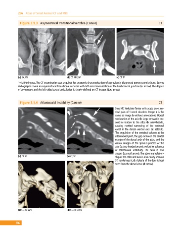
Figure 3.1.3 Asymmetrical Transitional Vertebra (Canine) CT
(a) DX, VD (b) CT, MIP, DP (c) CT, TP
1y M Pekingese. The CT examination was acquired for anatomic characterization of a previously diagnosed portosystemic shunt. Survey
radiographs reveal an asymmetrical transitional vertebra with left‐sided sacralization at the lumbosacral junction (a: arrow). The degree
of asymmetry and the left‐sided sacral articulation is clearly defined on CT images (b,c: arrow).
Figure 3.1.4 Atlantoaxial Instability (Canine) CT
5mo MC Yorkshire Terrier with acute onset cer
vical pain of 1‐week duration. Image a is the
same as image b without annotations. Dorsal
subluxation of the axis (b: large arrows) is pre
sent in relation to the atlas (b: arrowheads),
causing marked narrowing of the vertebral
canal in the dorsal–ventral axis (b: asterisk).
The angulation of the vertebral column at the
atlantoaxial joint, the gap between the caudal
margin of the dorsal arch of the atlas, and the
cranial margin of the spinous process of the
axis (b: two‐headed arrow) are further evidence
of atlantoaxial instability. The dens is also
absent (b: small arrow). The abnormal relation
(a) CT, SP (b) CT, SP ship of the atlas and axis is also clearly seen on
3D renderings (c,d). Aplasia of the dens is best
seen from the dorsal view (d: arrow).
(c) CT, 3D, LLAT (d) CT, 3D, DORS
286