Page 299 - Atlas of Small Animal CT and MRI
P. 299

Developmental Disorders  289


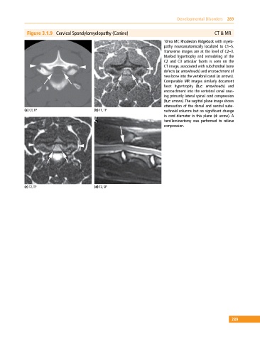
              Figure 3.1.9  Cervical Spondylomyelopathy (Canine)                                     CT & MR
                                                                             10mo MC Rhodesian Ridgeback with myelo­
                                                                             pathy neuroanatomically localized to C1–5.
                                                                             Transverse images are at the level of C2–3.
                                                                             Marked hypertrophy and remodeling of the
                                                                             C2  and  C3  articular facets  is  seen  on  the
                                                                             CT image, associated with subchondral bone
                                                                             defects (a: arrowheads) and encroachment of
                                                                             new bone into the vertebral canal (a: arrows).
                                                                             Comparable MR images similarly document
                                                                             facet hypertrophy (b,c: arrowheads) and
                                                                             encroachment into the vertebral canal caus­
                                                                             ing primarily lateral spinal cord compression
                                                                             (b,c: arrows). The sagittal plane image shows
                                                                             attenuation of the  dorsal and ventral suba­
            (a) CT, TP                       (b) T1, TP                      rachnoid columns but no significant change
                                                                             in cord diameter in this plane (d: arrow). A
                                                                             hemilaminectomy was performed to relieve
                                                                             compression.


            (c) T2, TP                       (d) T2, SP


                                                                                                             289
   294   295   296   297   298   299   300   301   302   303   304