
Page 302 - Atlas of Small Animal CT and MRI
P. 302
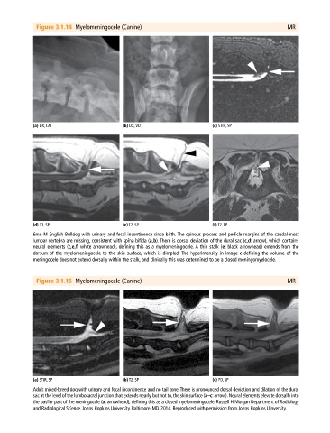
Figure 3.1.14  Myelomeningocele (Canine)                                                    MR


             (a) DX, LAT                      (b) DX, VD                       (c) STIR, SP


             (d) T1, SP                       (e) T2, SP                       (f) T2, TP
             6mo M English Bulldog with urinary and fecal incontinence since birth. The spinous process and pedicle margins of the caudal‐most
             lumbar vertebra are missing, consistent with spina bifida (a,b). There is dorsal deviation of the dural sac (c,d: arrow), which contains
             neural elements (c,e,f: white arrowhead), defining this as a myelomeningocele. A thin stalk (e: black arrowhead) extends from the
               dorsum of the myelomeningocele to the skin surface, which is dimpled. The hyperintensity in image c defining the volume of the
               meningocele does not extend dorsally within the stalk, and clinically this was determined to be a closed meningomyelocele.


              Figure 3.1.15  Myelomeningocele (Canine)                                                    MR


             (a) STIR, SP                     (b) T2, SP                       (c) PD, SP
             Adult mixed‐breed dog with urinary and fecal incontinence and no tail tone. There is pronounced dorsal deviation and dilation of the dural
             sac at the level of the lumbosacral junction that extends nearly, but not to, the skin surface (a–c: arrow). Neural elements elevate dorsally into
             the basilar part of the meningocele (a: arrowhead), defining this as a closed myelomeningocele. Russell H Morgan Department of Radiology
             and Radiological Science, Johns Hopkins University, Baltimore, MD, 2014. Reproduced with permission from Johns Hopkins Uinversity.