
Page 300 - Atlas of Small Animal CT and MRI
P. 300
290 Atlas of Small Animal CT and MRI
Figure 3.1.10 Cervical Spondylomyelopathy (Canine) MR
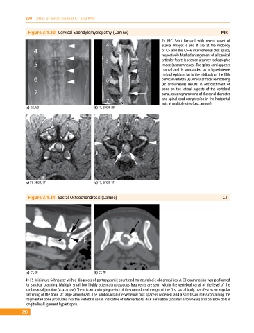
2y MC Saint Bernard with recent onset of
ataxia. Images c and d are at the midbody
of C5 and the C5–6 intervertebral disk space,
respectively. Marked enlargement of all cervical
articular facets is seen on a survey radiographic
image (a: arrowheads). The spinal cord appears
normal and is surrounded by a hyperintense
halo of epidural fat in the midbody of the fifth
cervical vertebra (c). Articular facet remodeling
(d: arrowheads) results in encroachment of
bone on the lateral aspects of the vertebral
canal, causing narrowing of the canal diameter
and spinal cord compression in the horizontal
axis at multiple sites (b,d: arrows).
(a) DX, VD (b) T1, SPGR, DP
(c) T1, SPGR, TP (d) T1, SPGR, TP
Figure 3.1.11 Sacral Osteochondrosis (Canine) CT
(a) CT, SP (b) CT, TP
4y FS Miniature Schnauzer with a diagnosis of portosystemic shunt and no neurologic abnormalities. A CT examination was performed
for surgical planning. Multiple small but highly attenuating osseous fragments are seen within the vertebral canal at the level of the
lumbosacral junction (a,b: arrow). There is an underlying defect of the craniodorsal margin of the first sacral body, manifest as an angular
flattening of the bone (a: large arrowhead). The lumbosacral intervertebral disk space is widened, and a soft‐tissue mass containing the
fragmented bone protrudes into the vertebral canal, indicative of intervertebral disk herniation (a: small arrowhead) and possible dorsal
longitudinal ligament hypertrophy.
290