
Page 297 - Atlas of Small Animal CT and MRI
P. 297
Developmental Disorders 287
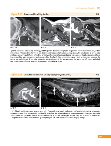
Figure 3.1.5 Atlantoaxial Instability (Canine) MR
(a) DX, LAT (b) T1, SP (c) T2, SP
7y FS Maltese with a 4‐day history of lethargy and tetraparesis. The survey radiographic image shows a complex craniocervical junction
malformation with occipital malformation and abnormal atlantooccipital articulation (a: large arrow), hypoplastic dens (a: arrowhead),
misshapen axis with caudal fusion to the third cervical vertebra (a: small arrow), and dorsal subluxation of the atlantoaxial joint. There
is widening of the space between the caudal margin of the dorsal arch of the atlas and the cranial margin of the spinous process of the
axis (a: two‐headed arrow). Atlantoaxial subluxation and dens hypoplasia (b,c: arrowhead) are also seen on the MR images, and spinal
cord compression can be seen at the site of atlantoaxial subluxation (c: arrow).
Figure 3.1.6 Chiari‐like Malformation with Syringohydromyelia (Canine) MR
(a) T2, SP (b) T2, SP
1.5y F Chihuahua with recent‐onset paroxysmal episodes. The caudal cranial vault is small as a result of occipital hypoplasia (a: arrowhead),
and moderate generalized hydrocephalus is evident (a). Marked saccular syringohydromyelia is present throughout the cervical and cranial
thoracic spinal cord (b: arrows). There is also T2 hyperintensity within cord parenchyma, which is likely due to edema (b: arrowhead).
A diagnosis of Chiari‐like malformation with syringohydromyelia was made based on clinical and imaging findings.
287