
Page 301 - Atlas of Small Animal CT and MRI
P. 301
Developmental Disorders 291
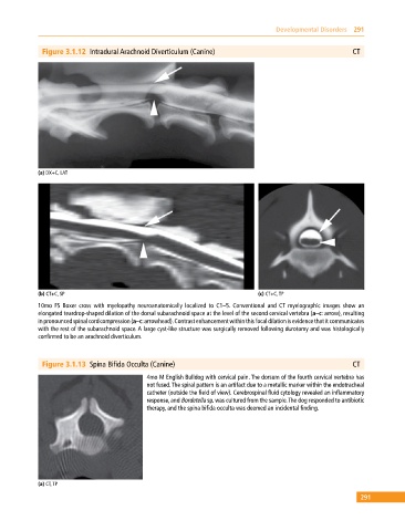
Figure 3.1.12 Intradural Arachnoid Diverticulum (Canine) CT
(a) DX+C, LAT
(b) CT+C, SP (c) CT+C, TP
10mo FS Boxer cross with myelopathy neuroanatomically localized to C1–5. Conventional and CT myelographic images show an
elongated teardrop‐shaped dilation of the dorsal subarachnoid space at the level of the second cervical vertebra (a–c: arrow), resulting
in pronounced spinal cord compression (a–c: arrowhead). Contrast enhancement within this focal dilation is evidence that it communicates
with the rest of the subarachnoid space. A large cyst‐like structure was surgically removed following durotomy and was histologically
confirmed to be an arachnoid diverticulum.
Figure 3.1.13 Spina Bifida Occulta (Canine) CT
4mo M English Bulldog with cervical pain. The dorsum of the fourth cervical vertebra has
not fused. The spiral pattern is an artifact due to a metallic marker within the endotracheal
catheter (outside the field of view). Cerebrospinal fluid cytology revealed an inflammatory
response, and Bordetella sp. was cultured from the sample. The dog responded to antibiotic
therapy, and the spina bifida occulta was deemed an incidental finding.
(a) CT, TP
291