
Page 473 - Atlas of Small Animal CT and MRI
P. 473
Small Airways and Parenchyma 463
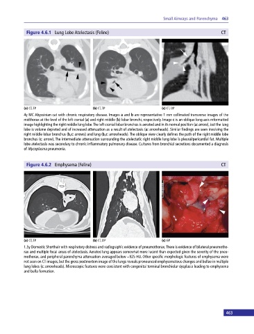
Figure 4.6.1 Lung Lobe Atelectasis (Feline) CT
(a) CT, TP (b) CT, TP (c) CT, OP
4y MC Abyssinian cat with chronic respiratory disease. Images a and b are representative 1 mm collimated transverse images of the
midthorax at the level of the left cranial (a) and right middle (b) lobar bronchi, respectively. Image c is an oblique long‐axis reformatted
image highlighting the right middle lung lobe. The left cranial lobar bronchus is aerated and in its normal position (a: arrow), but the lung
lobe is volume depleted and of increased attenuation as a result of atelectasis (a: arrowheads). Similar findings are seen involving the
right middle lobar bronchus (b,c: arrows) and lung (b,c: arrowheads). The oblique view clearly defines the path of the right middle lobe
bronchus (c: arrow). The intermediate attenuation surrounding the atelectatic right middle lung lobe is pleural/pericardial fat. Multiple
lobe atelectasis was secondary to chronic inflammatory pulmonary disease. Cultures from bronchial secretions documented a diagnosis
of Mycoplasma pneumonia.
Figure 4.6.2 Emphysema (Feline) CT
(a) CT, TP (b) CT, DP (c) GP
1.5y Domestic Shorthair with respiratory distress and radiographic evidence of pneumothorax. There is evidence of bilateral pneumotho-
rax and multiple focal areas of atelectasis. Aerated lung appears somewhat more lucent than expected given the severity of the pneu-
mothorax, and peripheral parenchyma attenuation averaged below −925 HU. Other specific morphologic features of emphysema were
not seen on CT images, but the gross postmortem image of the lungs reveals pronounced emphysematous changes and bullae in multiple
lung lobes (c: arrowheads). Microscopic features were consistent with congenital terminal bronchiolar dysplasia leading to emphysema
and bulla formation.
463