
Page 475 - Atlas of Small Animal CT and MRI
P. 475
Small Airways and Parenchyma 465
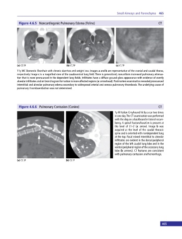
Figure 4.6.5 Noncardiogenic Pulmonary Edema (Feline) CT
(a) CT, TP (b) CT, TP (c) CT, TP
11y MC Domestic Shorthair with chronic diarrhea and weight loss. Images a and b are representative of the cranial and caudal thorax,
respectively. Image c is a magnified view of the caudoventral lung field. There is generalized, nonuniform increased pulmonary attenua-
tion that is more pronounced in the dependent lung fields. Infiltrates have a diffuse ground‐glass appearance with evidence of overtly
alveolar infiltrates and air bronchogram formation in more affected regions (a: arrowhead). Postmortem examination revealed pronounced
interstitial and alveolar pulmonary edema secondary to widespread arterial and venous pulmonary thrombosis. The underlying cause of
pulmonary thromboembolism was not determined.
Figure 4.6.6 Pulmonary Contusion (Canine) CT
1y M Italian Greyhound hit by a car two times
in one day. The CT examination was performed
with the dog on a backboard in lateral recum-
bency. A spinal fracture/luxation is present at
the level of L1–2 (a: arrow). Image b was
acquired at the level of the caudal thoracic
spine and is oriented with nondependent lung
at the top. Focal mixed interstitial to alveolar
infiltrates are evident in the dorsal peripheral
region of the left caudal lung lobe and in the
ventral peripheral region of the accessory lung
lobe (b: arrows). CT features are consistent
with pulmonary contusion and hemorrhage.
(a) CT, SP (b) CT, TP
465