
Page 478 - Atlas of Small Animal CT and MRI
P. 478
468 Atlas of Small Animal CT and MRI
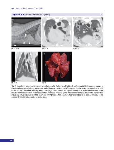
Figure 4.6.9 Interstitial Pneumonia (Feline) CT
(a) DX, RLAT (b) CT, TP (c) CT, TP
(d) CT, DP (e) GP
12y FS Ragdoll with progressive respiratory signs. Radiographic findings include diffuse bronchointerstitial infiltrates that coalesce to
alveolar infiltrates ventrally (a: arrowheads) and marked bronchiectasis (a: arrow). CT images confirm the presence of generalized bronchi-
ectasis and alveolar infiltrates involving the left cranial, right cranial, and left and right caudal lung lobes (b–d). Bronchoalveolar lavage
revealed moderate suppurative inflammation without evidence of infectious agents. Postmortem examination documented bronchiectasis
and severe, diffuse and acute interstitial pneumonia with fibrin exudation, alveolar histiocytosis, and septal fibrosis (e). Infectious agents
were not detected on either routine or special stains.
468