
Page 476 - Atlas of Small Animal CT and MRI
P. 476
466 Atlas of Small Animal CT and MRI
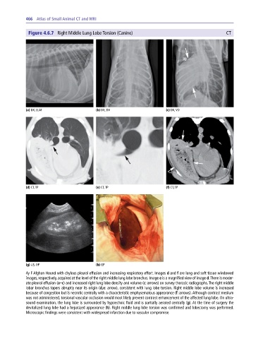
Figure 4.6.7 Right Middle Lung Lobe Torsion (Canine) CT
(a) DX, LLAT (b) DX, DV (c) DX, VD
(d) CT, TP (e) CT, TP (f) CT, TP
(g) US, OP (h) GP
4y F Afghan Hound with chylous pleural effusion and increasing respiratory effort. Images d and f are lung and soft‐tissue windowed
images, respectively, acquired at the level of the right middle lung lobe bronchus. Image e is a magnified view of image d. There is moder-
ate pleural effusion (a–c) and increased right lung lobe density and volume (c: arrows) on survey thoracic radiographs. The right middle
lobar bronchus tapers abruptly near its origin (d,e: arrow), consistent with lung lobe torsion. Right middle lobe volume is increased
because of congestion but is necrotic centrally with a characteristic emphysematous appearance (f: arrows). Although contrast medium
was not administered, torsional vascular occlusion would most likely prevent contrast enhancement of the affected lung lobe. On ultra-
sound examination, the lung lobe is surrounded by hypoechoic fluid and is partially aerated centrally (g). At the time of surgery the
devitalized lung lobe had a hepatized appearance (h). Right middle lung lobe torsion was confirmed and lobectomy was performed.
Microscopic findings were consistent with widespread infarction due to vascular compromise.