
Page 643 - Atlas of Small Animal CT and MRI
P. 643
Developmental and Metabolic Disorders 633
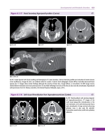
Figure 6.1.17 Renal Secondary Hyperparathyroidism (Canine) CT
(a) DX, LAT (b) CT, TP (c) CT, TP
(d) CT, MIP, LAT
9y MC Cocker Spaniel with facial swelling and hemoptysis of 3‐week duration. Clinical chemistry profile was indicative of severe chronic
renal insufficiency. Images b and c are ordered rostral to caudal. A lateral skull radiograph reveals diffuse markedly decreased bone
opacity (a). CT images show similar findings although the severity of bone loss, though diffuse, is nonuniform. An MIP image reveals
skeletal demineralization to be most pronounced in the skull (d). Pathologic fractures of the ribs are also seen (d: arrowheads). Reproduced
with permission from Dr. Shimizu Junichiro, Uni Animal Hospital, Hokkaido, Japan, 2014.
Figure 6.1.18 Soft‐tissue Mineralization from Hyperadrenocorticism (Canine) CT
8y MC Newfoundland with pituitary‐depend
ent hyperadrenocorticism. CT images of the
skull reveal plaque‐like mineralization in the
skin (calcinosis cutis) and intermuscular fascia
(a,b: arrowheads). A large contrast‐enhancing
pituitary mass is also seen (b: asterisk).
Postmortem examination confirmed a pituitary
adenoma.
(a) CT+C, TP (b) CT+C, TP
633