
Page 640 - Atlas of Small Animal CT and MRI
P. 640
630 Atlas of Small Animal CT and MRI
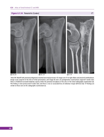
Figure 6.1.14 Panosteitis (Canine) CT
(a) DX, LAT (b) DX, LAT (c) CT, SP
14mo MC Mastiff with previously diagnosed ununited anconeal processes. All images are of the right elbow and proximal antebrachium.
Image a was acquired at the time of initial presentation, and images b and c are postoperative examinations acquired 6 weeks later.
There is ill‐defined increased medullary opacity within the proximal metaphysis of the ulna on the initial radiographic examination (a).
This finding is more pronounced 6 weeks later, and there is now an associated loss of endosteal margin definition (b). CT findings are
similar to those seen on the radiographic examination (c).
630