
Page 636 - Atlas of Small Animal CT and MRI
P. 636
626 Atlas of Small Animal CT and MRI
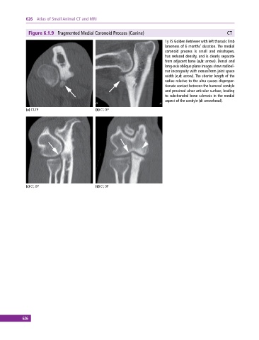
Figure 6.1.9 Fragmented Medial Coronoid Process (Canine) CT
1y FS Golden Retriever with left thoracic limb
lameness of 6 months’ duration. The medial
coronoid process is small and misshapen,
has reduced density, and is clearly separate
from adjacent bone (a,b: arrow). Dorsal and
long‐axis oblique plane images show radioul
nar incongruity with nonuniform joint space
width (c,d: arrow). The shorter length of the
radius relative to the ulna causes dispropor
tionate contact between the humeral condyle
and proximal ulnar articular surface, leading
to subchondral bone sclerosis in the medial
aspect of the condyle (d: arrowhead).
(a) CT, TP (b) CT, OP
(c) CT, OP (d) CT, DP
626