
Page 637 - Atlas of Small Animal CT and MRI
P. 637
Developmental and Metabolic Disorders 627
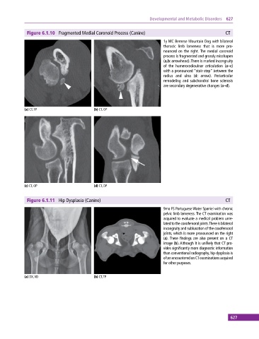
Figure 6.1.10 Fragmented Medial Coronoid Process (Canine) CT
1y MC Bernese Mountain Dog with bilateral
thoracic limb lameness that is more pro
nounced on the right. The medial coronoid
process is fragmented and grossly misshapen
(a,b: arrowhead). There is marked incongruity
of the humeroradioulnar articulation (a–c)
with a pronounced “stair‐step” between the
radius and ulna (d: arrow). Periarticular
remodeling and subchondral bone sclerosis
are secondary degenerative changes (a–d).
(a) CT, TP (b) CT, OP
(c) CT, OP (d) CT, DP
Figure 6.1.11 Hip Dysplasia (Canine) CT
9mo FS Portuguese Water Spaniel with chronic
pelvic limb lameness. The CT examination was
acquired to evaluate a medical problem unre
lated to the coxofemoral joints. There is bilateral
incongruity and subluxation of the coxofemoral
joints, which is more pronounced on the right
(a). These findings are also present on a CT
image (b). Although it is unlikely that CT pro
vides significantly more diagnostic information
than conventional radiography, hip dysplasia is
often encountered on CT examinations acquired
for other purposes.
(a) DX, VD (b) CT, TP
627