
Page 633 - Atlas of Small Animal CT and MRI
P. 633
Developmental and Metabolic Disorders 623
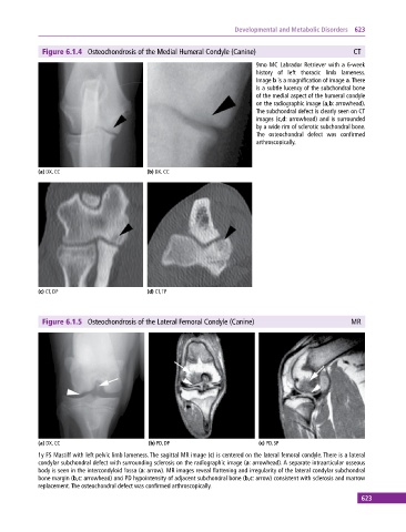
Figure 6.1.4 Osteochondrosis of the Medial Humeral Condyle (Canine) CT
9mo MC Labrador Retriever with a 6‐week
history of left thoracic limb lameness.
Image b is a magnification of image a. There
is a subtle lucency of the subchondral bone
of the medial aspect of the humeral condyle
on the radiographic image (a,b: arrowhead).
The subchondral defect is clearly seen on CT
images (c,d: arrowhead) and is surrounded
by a wide rim of sclerotic subchondral bone.
The osteochondral defect was confirmed
arthroscopically.
(a) DX, CC (b) DX, CC
(c) CT, DP (d) CT, TP
Figure 6.1.5 Osteochondrosis of the Lateral Femoral Condyle (Canine) MR
(a) DX, CC (b) PD, DP (c) PD, SP
1y FS Mastiff with left pelvic limb lameness. The sagittal MR image (c) is centered on the lateral femoral condyle. There is a lateral
condylar subchondral defect with surrounding sclerosis on the radiographic image (a: arrowhead). A separate intraarticular osseous
body is seen in the intercondyloid fossa (a: arrow). MR images reveal flattening and irregularity of the lateral condylar subchondral
bone margin (b,c: arrowhead) and PD hypointensity of adjacent subchondral bone (b,c: arrow) consistent with sclerosis and marrow
replacement. The osteochondral defect was confirmed arthroscopically.
623