
Page 632 - Atlas of Small Animal CT and MRI
P. 632
622 Atlas of Small Animal CT and MRI
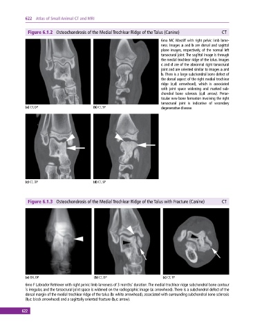
Figure 6.1.2 Osteochondrosis of the Medial Trochlear Ridge of the Talus (Canine) CT
6mo MC Mastiff with right pelvic limb lame
ness. Images a and b are dorsal and sagittal
plane images, respectively, of the normal left
tarsocrural joint. The sagittal image is through
the medial trochlear ridge of the talus. Images
c and d are of the abnormal right tarsocrural
joint and are oriented similar to images a and
b. There is a large subchondral bone defect of
the dorsal aspect of the right medial trochlear
ridge (c,d: arrowhead), which is associated
with joint space widening and marked sub
chondral bone sclerosis (c,d: arrow). Periar
ticular new bone formation involving the right
tarsocrural joint is indicative of secondary
(a) CT, DP (b) CT, SP degenerative disease.
(c) CT, DP (d) CT, SP
Figure 6.1.3 Osteochondrosis of the Medial Trochlear Ridge of the Talus with Fracture (Canine) CT
(a) DX, DP (b) CT, DP (c) CT, TP
6mo F Labrador Retriever with right pelvic limb lameness of 3 months’ duration. The medial trochlear ridge subchondral bone contour
is irregular, and the tarsocrural joint space is widened on the radiographic image (a: arrowhead). There is a subchondral defect of the
dorsal margin of the medial trochlear ridge of the talus (b: white arrowhead), associated with surrounding subchondral bone sclerosis
(b,c: black arrowhead) and a sagittally oriented fracture (b,c: arrow).
622