
Page 631 - Atlas of Small Animal CT and MRI
P. 631
Developmental and Metabolic Disorders 621
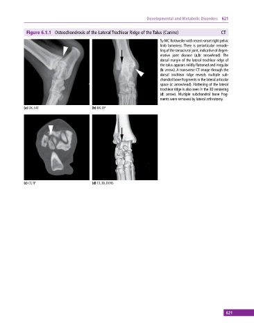
Figure 6.1.1 Osteochondrosis of the Lateral Trochlear Ridge of the Talus (Canine) CT
5y MC Rottweiler with recent‐onset right pelvic
limb lameness. There is periarticular remode
ling of the tarsocrural joint, indicative of degen
erative joint disease (a,b: arrowhead). The
dorsal margin of the lateral trochlear ridge of
the talus appears mildly flattened and irregular
(b: arrow). A transverse CT image through the
dorsal trochlear ridge reveals multiple sub
chondral bone fragments in the lateral articular
space (c: arrowhead). Flattening of the lateral
trochlear ridge is also seen in the 3D rendering
(d: arrow). Multiple subchondral bone frag
ments were removed by lateral arthrotomy.
(a) DX, LAT (b) DX, DP
(c) CT, TP (d) CT, 3D, DORS
621