
Page 635 - Atlas of Small Animal CT and MRI
P. 635
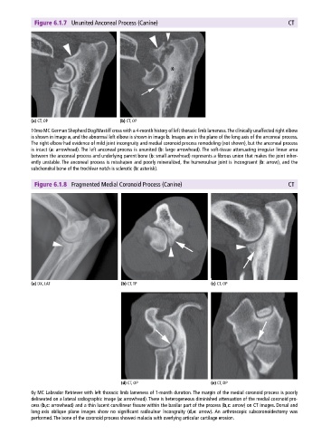
Figure 6.1.7 Ununited Anconeal Process (Canine) CT
(a) CT, OP (b) CT, OP
10mo MC German Shepherd Dog/Mastiff cross with a 4‐month history of left thoracic limb lameness. The clinically unaffected right elbow
is shown in image a, and the abnormal left elbow is shown in image b. Images are in the plane of the long axis of the anconeal process.
The right elbow had evidence of mild joint incongruity and medial coronoid process remodeling (not shown), but the anconeal process
is intact (a: arrowhead). The left anconeal process is ununited (b: large arrowhead). The soft‐tissue attenuating irregular linear area
between the anconeal process and underlying parent bone (b: small arrowhead) represents a fibrous union that makes the joint inher
ently unstable. The anconeal process is misshapen and poorly mineralized, the humeroulnar joint is incongruent (b: arrow), and the
subchondral bone of the trochlear notch is sclerotic (b: asterisk).
Figure 6.1.8 Fragmented Medial Coronoid Process (Canine) CT
(a) DX, LAT (b) CT, TP (c) CT, OP
(d) CT, OP (e) CT, DP
6y MC Labrador Retriever with left thoracic limb lameness of 1‐month duration. The margin of the medial coronoid process is poorly
delineated on a lateral radiographic image (a: arrowhead). There is heterogeneous diminished attenuation of the medial coronoid pro
cess (b,c: arrowhead) and a thin lucent curvilinear fissure within the basilar part of the process (b,c: arrow) on CT images. Dorsal and
long‐axis oblique plane images show no significant radioulnar incongruity (d,e: arrow). An arthroscopic subcoronoidectomy was
performed. The bone of the coronoid process showed malacia with overlying articular cartilage erosion.