
Page 634 - Atlas of Small Animal CT and MRI
P. 634
624 Atlas of Small Animal CT and MRI
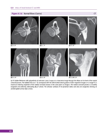
Figure 6.1.6 Normal Elbow (Canine) CT
(a) CT, TP (b) CT, TP (c) CT, OP
(d) CT, OP (e) CT, DP (f) CT, MIP, OP
4y FS Golden Retriever with polyarthritis of unknown cause. Image a is a transverse image through the elbow at the level of the medial
coronoid process. The labeled lines (a: C–E) correspond with the reformatted viewing plane of their respective images c–e. Image f is a
maximum‐intensity projection of the medial coronoid process in the same plane as image c. The medial coronoid process is smoothly
margined and uniformly attenuating (b,c,f: arrow). The articular surfaces of the proximal radius and ulna are congruent, forming an
uninterrupted surface (d,e: arrow).
624