
Page 639 - Atlas of Small Animal CT and MRI
P. 639
Developmental and Metabolic Disorders 629
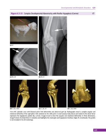
Figure 6.1.13 Complex Developmental Abnormality with Patellar Hypoplasia (Canine) CT
(a) XC
(c) DX, LAT (b) DX, CC
(d) CT, 3D, CRAN (e) CT, 3D, LAT (f) CT, 3D, CRAN
10mo MC Labrador cross with bilateral pelvic limb deformities and abnormal gait (a). Radiographs reveal a complex angular and
rotational deformity of the right pelvic limb centered on the stifle joint. A small osseous body dorsal and medial to the distal femur
represents the hypoplastic patella (b,c: arrow). Images d and e show the angular and rotational deformities in three dimensions.
Image f shows the distal femur in isolation and highlights the misshapen and hypoplastic trochlear ridges (f: arrowheads). The patella
is not included in the 3D renderings.
629