
Page 686 - Atlas of Small Animal CT and MRI
P. 686
676 Atlas of Small Animal CT and MRI
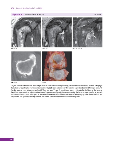
Figure 6.5.1 Osteoarthritis (Canine) CT & MR
(a) T1, SP (b) T2, SP (c) T1+C+FS, SP
(d) CT, TP (e) GP
10y MC Golden Retriever with chronic right thoracic limb lameness and previously performed biceps tenectomy. There is osteophyte
formation surrounding the humerus and glenoid cavity (a,b: open arrowhead). This is better appreciated on the CT images surround-
ing the humeral head (d: open arrowheads). There is a focal T1 and T2 hypointense region in the subchondral bone of the humeral
head (a,b: open arrow), which contrast enhances (c: open arrow). The soft tissues surrounding the humerus are enhancing (c: arrows),
and the void in the caudal joint space (c: arrowhead) represents joint effusion with a rim of enhancing synovial tissue. The limb was
amputated, and synovitis, cartilage erosion, and severe osteoarthritis were confirmed histologically.
676