
Page 687 - Atlas of Small Animal CT and MRI
P. 687
Degenerative Disorders 677
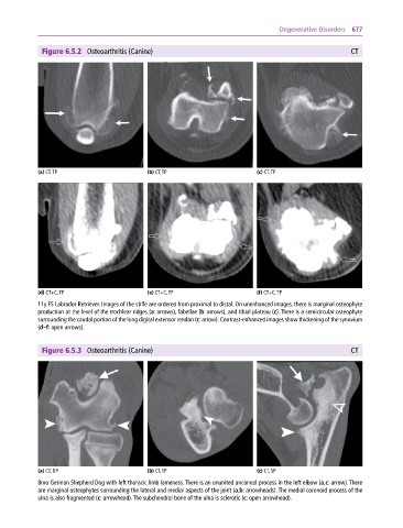
Figure 6.5.2 Osteoarthritis (Canine) CT
(a) CT, TP (b) CT, TP (c) CT, TP
(d) CT+C, TP (e) CT+C, TP (f) CT+C, TP
11y FS Labrador Retriever. Images of the stifle are ordered from proximal to distal. On unenhanced images, there is marginal osteophyte
production at the level of the trochlear ridges (a: arrows), fabellae (b: arrows), and tibial plateau (c). There is a semicircular osteophyte
surrounding the caudal portion of the long digital extensor tendon (c: arrow). Contrast‐enhanced images show thickening of the synovium
(d–f: open arrows).
Figure 6.5.3 Osteoarthritis (Canine) CT
(a) CT, DP (b) CT, TP (c) CT, SP
8mo German Shepherd Dog with left thoracic limb lameness. There is an ununited anconeal process in the left elbow (a,c: arrow). There
are marginal osteophytes surrounding the lateral and medial aspects of the joint (a,b: arrowheads). The medial coronoid process of the
ulna is also fragmented (c: arrowhead). The subchondral bone of the ulna is sclerotic (c: open arrowhead).