Page 688 - Atlas of Small Animal CT and MRI
P. 688

678  Atlas of Small Animal CT and MRI


              Figure 6.5.4  Synovitis (Feline)                                                            MR
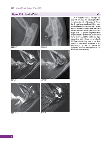
                                                                              5y MC Domestic Mediumhair with right tho-
                                                                              racic limb lameness. On radiographs of the
                                                                              elbow (a,b), there is mild osteophyte forma-
                                                                              tion (b: small arrow) and medial joint space
                                                                              widening (b: open arrowhead), which is most
                                                                              apparent on the craniocaudal projection. There
                                                                              is marked distension of the elbow joint on MR
                                                                              images (c–f). The synovial component of the
                                                                              joint distension is predominant on enhanced
                                                                              images (e: arrow), with the central low signal
                                                                              representing joint effusion (e: arrowhead).
                                                                              STIR hyperintensity is present in the distal
                                                                              humerus (f: open arrow). A diagnosis of lym-
                                                                              phoplasmacytic  synovitis  and  synovial  cell
             (a) DX, LAT                      (b) DX, CC                      hyperplasia associated with degenerative joint
                                                                              disease was made on biopsy.


             (c) T2, SP                       (d) PD, SP


             (e) T1+C, SP                     (f) ST, SP


           678
   683   684   685   686   687   688   689   690   691   692   693