
Page 683 - Atlas of Small Animal CT and MRI
P. 683
Neoplasia 673
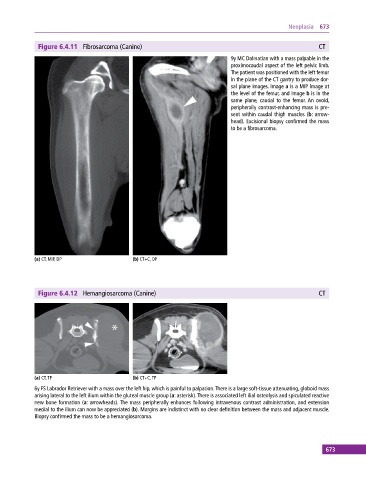
Figure 6.4.11 Fibrosarcoma (Canine) CT
9y MC Dalmatian with a mass palpable in the
proximocaudal aspect of the left pelvic limb.
The patient was positioned with the left femur
in the plane of the CT gantry to produce dor-
sal plane images. Image a is a MIP image at
the level of the femur, and image b is in the
same plane, caudal to the femur. An ovoid,
peripherally contrast‐enhancing mass is pre-
sent within caudal thigh muscles (b: arrow-
head). Excisional biopsy confirmed the mass
to be a fibrosarcoma.
(a) CT, MIP, DP (b) CT+C, DP
Figure 6.4.12 Hemangiosarcoma (Canine) CT
(a) CT, TP (b) CT+C, TP
6y FS Labrador Retriever with a mass over the left hip, which is painful to palpation. There is a large soft‐tissue attenuating, globoid mass
arising lateral to the left ilium within the gluteal muscle group (a: asterisk). There is associated left ilial osteolysis and spiculated reactive
new bone formation (a: arrowheads). The mass peripherally enhances following intravenous contrast administration, and extension
medial to the ilium can now be appreciated (b). Margins are indistinct with no clear definition between the mass and adjacent muscle.
Biopsy confirmed the mass to be a hemangiosarcoma.
673