
Page 678 - Atlas of Small Animal CT and MRI
P. 678
668 Atlas of Small Animal CT and MRI
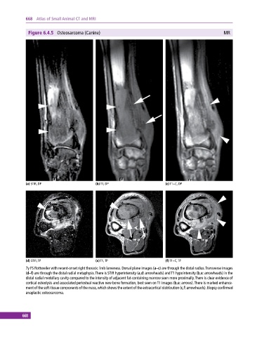
Figure 6.4.5 Osteosarcoma (Canine) MR
(a) STIR, DP (b) T1, DP (c) T1+C, DP
(d) STIR, TP (e) T1, TP (f) T1+C, TP
7y FS Rottweiler with recent‐onset right thoracic limb lameness. Dorsal plane images (a–c) are through the distal radius. Transverse images
(d–f) are through the distal radial metaphysis. There is STIR hyperintensity (a,d: arrowheads) and T1 hypointensity (b,e: arrowheads) in the
distal radial medullary cavity compared to the intensity of adjacent fat‐containing marrow seen more proximally. There is clear evidence of
cortical osteolysis and associated periosteal reactive new bone formation, best seen on T1 images (b,e: arrows). There is marked enhance-
ment of the soft‐tissue components of the mass, which shows the extent of the extracortical distribution (c,f: arrowheads). Biopsy confirmed
anaplastic osteosarcoma.
668