
Page 674 - Atlas of Small Animal CT and MRI
P. 674
664 Atlas of Small Animal CT and MRI
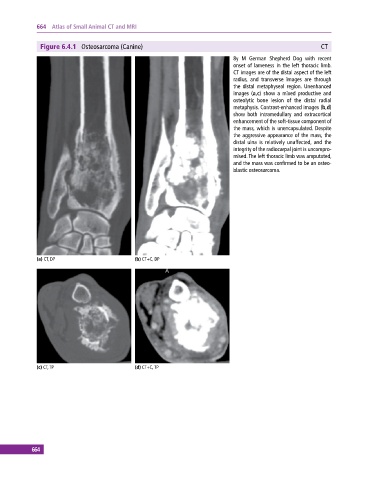
Figure 6.4.1 Osteosarcoma (Canine) CT
8y M German Shepherd Dog with recent
onset of lameness in the left thoracic limb.
CT images are of the distal aspect of the left
radius, and transverse images are through
the distal metaphyseal region. Unenhanced
images (a,c) show a mixed productive and
osteolytic bone lesion of the distal radial
metaphysis. Contrast‐enhanced images (b,d)
show both intramedullary and extracortical
enhancement of the soft‐tissue component of
the mass, which is unencapsulated. Despite
the aggressive appearance of the mass, the
distal ulna is relatively unaffected, and the
integrity of the radiocarpal joint is uncompro-
mised. The left thoracic limb was amputated,
and the mass was confirmed to be an osteo-
blastic osteosarcoma.
(a) CT, DP (b) CT+C, DP
(c) CT, TP (d) CT+C, TP
664