
Page 669 - Atlas of Small Animal CT and MRI
P. 669
Inflammatory Disorders 659
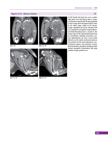
Figure 6.3.6 Abscess (Canine) CT
6y MC Mastiff with high fever and a swollen
right pelvic limb with pitting edema. Images
a and b were acquired with the dog positioned
so that images show the proximal pelvic limbs
in the dorsal plane caudal to the femurs.
Image c represents the normal left pelvic limb
as a comparison to image d. A large multicam-
eral fluid‐attenuating mass is present in the
caudal aspect of the right proximal pelvic limb
(a: arrowheads). Following intravenous con-
trast administration, the mass is more clearly
defined as a thin‐walled, multicameral abscess
that extends into the semimembranosus, sem-
itendinosus, gluteal, and quadriceps muscles
(a) CT, DP (b) CT+C, DP (b,d: arrowheads). Aspiration cytology revealed
marked neutrophilic inflammation with large
numbers of gram‐positive cocci.
(c) CT+C, SP (d) CT+C, SP
659