
Page 668 - Atlas of Small Animal CT and MRI
P. 668
658 Atlas of Small Animal CT and MRI
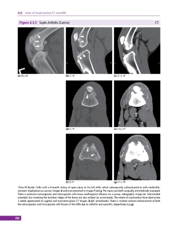
Figure 6.3.5 Septic Arthritis (Canine) CT
(a) DX, LAT (b) CT, SP (c) CT+C, SP
(d) CT, TP (e) CT+C, TP
(f) CT, TP (g) CT+C, TP
15mo M Border Collie with a 6‐month history of open injury to the left stifle, which subsequently cultured positive with methicillin‐
resistant Staphylococcus aureus. Images d and e are proximal to images f and g. The injury was both surgically and medically managed.
There is extensive extracapsular and intracapsular soft‐tissue swelling/joint effusion on a survey radiographic image (a). Subchondral
osteolytic foci involving the trochlear ridges of the femur are also evident (a: arrowheads). The extent of subchondral bone destruction
is better appreciated on sagittal and transverse plane CT images (b,d,f: arrowheads). There is marked contrast enhancement of both
the extracapsular and intracapsular soft tissues of the stifle due to cellulitis and synovitis, respectively (c,e,g).
658