
Page 667 - Atlas of Small Animal CT and MRI
P. 667
Inflammatory Disorders 657
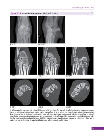
Figure 6.3.4 Erosive Immune‐mediated Polyarthritis (Canine) CT
(a) DX, DP (b) DX, LAT
(c) CT, DP (d) CT, DP (e) CT, DP
(f) CT, TP (g) CT, TP (h) CT, TP
8y MC Labrador Retriever cross with a 3‐month history of pelvic limb discomfort and right carpal valgus deviation. A pain response was
elicited on palpation of multiple joints. Images c–e are ordered from dorsal to palmar, and images f–h are ordered from proximal to
distal. Radiographs of the right carpus reveal peri‐arthrodial soft‐tissue swelling and multiple osteolytic foci of the carpal and metacarpal
bones. Similar radiographic abnormalities were seen on radiographs of the left carpus. CT images reveal pronounced widespread sub-
chondral bone osteolysis. Cytology of synovial fluid from multiple joints revealed moderate suppurative inflammation. There was a
marked improvement in clinical signs and joint fluid cytology following steroid administration.
657